
25 Mar To Best Serve Your Patients, Protect Yourself First
Are cardiologists ready to combat COVID-19?
Doctors at Milan (Italy) L. Sacco Hospital to use new imaging technologies in the fight against coronavirus
In March 6, 2020 The American College of Cardiologist published the following bulletin
highlighting the importance of CV diagnostics in patients with COVID-19 infection, stressing the
need for echocardiographic investigation and the importance of using capable devices to counteract
the spreading of the infection.

To best serve your patients, protect yourself first! ……..
Decontaminate surfaces including stethoscope, cellular phones, computer peripherals, and other devices frequently.
- Cardiologists should be prepared to assist other clinical specialties in managing cardiac complications in severe cases of COVID-19
- Patients with underlying cardiovascular disease are at higher risk of contracting COVID-19 and have a worse prognosis
- Patients demonstrating heart failure, arrhythmia, ECG changes or cardiomegaly should have echocardiography
Despite this, the need for absolute isolation of symptomatic Patients and the high possibility of spreading the infection outside the isolation rooms in the High Intensity Care Departments, makes any attempt at an instrumental diagnostic approach difficult, given also the absolute need to decontaminate the equipment used after each individual examination. Echocardiography is the
most used diagnostic method for the evaluation of the cardiac function and more recently its use in the evaluation of the pulmonary parenchyma has reduced the use of radiographic investigations in areas of high care intensity. The advantages deriving from its possible execution at the patient’s bedside are well known but the high risk of serious coronavirus infection diffusion has made its
use difficult so far.
Disinfection is the only way to counter the risk of spreading the disease from one patient to another or among the operators themselves, which event is even more dangerous because it would jeopardize the tightness of the system.
Hand-carried (HC) echocardiography devices offer rapid and readily available information at the bedside, as they help overcome the problems caused by the use of cumbersome standard equipment.
The use of “ultramobile devices” has recently been introduced in the area of cardiovascular ultrasound diagnostics. These devices are miniaturized systems equipped with diagnostic quality two-dimensional (2D) and colour Doppler imaging. With simplified criteria they can detect left ventricular disfunction and moderate-severe valvulopathy with good sensitivity and specificity. They are extremely useful in the immediate diagnosis of pericardial effusion, of serious pathologies of the ascending thoracic aorta and in the definition of states of cardio-circulatory failure or inflammatory states of the pulmonary parenchyma. In resource-limited settings, ultra mobile systems can reduce the need for standard echocardiography, starting from these considerations and combining the professional resources in the Cardiology Department (director Dott. M.Viecca) and in the Anesthesia and Resuscitation Department
(director Dott. M.Catena) at the Luigi Sacco Hospital in Milan, in collaboration with a leading ultrasound sector company, a strategic intervention plan was designed for the performance of bedside echocardiographic examinations in COVID-19 patients, intubated and mechanically ventilated, admitted to the intensive care unit. The team includes Dr. Andrea Perotti (Anesthesia and Resuscitation Department), the Doctors of the Echocardiography Laboratory and the General Resuscitation Department.
The technology used is the Lumify platform (Philips Ultrasound), an “Ultramobile” system, offering a high level of integration of the equipment, with a dimension slightly larger than a common echocardiographic probe (fig. 1).
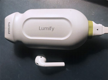
(Fig.1 The core of the Lumix system, enclosed in a probe with a high level of miniaturization)
The probe can be connected to any handheld device, smartphone or tablet (fig. 2), allowing the execution of cardiac, pulmonary, vascular, abdominal, soft tissue and joint ultrasound. The smart device is also a source of energy, with up to 3 hours of autonomy.
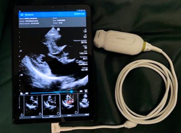
(Fig.2 The probe can be connected to any handheld device, smartphone or ,as in this case, tablet.)
The isolation system, required to avoid the danger of spreading the infection, is simple, consisting of a mono use coating sheath commonly used for picardial echocardiography in the CCH operating room. At the end of the examination, the probe is removed from the casing, reinserted in a new protective sheath and ready for a subsequent examination (fig. 3).
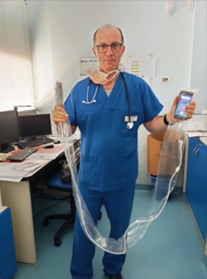
Fig.3 The compactness of the system makes it easily isolable from the environment with a mono use coating sheath, making it very practical in environments with a high level of contamination or where absolute sterility is required.
Thanks to internet connectivity, the images, stored in DICOM format, can also be simultaneously viewed in real time by other doctors outside the contaminated area. Finally, a video call system allows communication between operators. This aspect is decisive in the hypothesis of field Hospital installations, as it gives the possibility to exploit few medical imaging experts for the interpretation
of examinations from numerous Spot Centers, also performed by Doctors or trained non-medical staff not specialized in imaging.
This is a great organizational effort whose realization was made possible only by combining the professionalism of our Doctors with industrial know-how. Special thanks to all the benefactors whose donations have enabled our Center, among the leading institutions in the field worldwide, to develop such advanced technology which will allow us to offer patients the high quality care that our “Sistema Italia” has always offered.
…..Dilegua o notte, tramontate stelle, all’alba vincerò
Da Turandot
Alberto Barosi MD
Head of Cardiovascular Imaging Laboratory
Luigi Sacco Hospital, Milan